Sense Organs Quiz Questions And Answers

You must have learned about sense organs in primary school. But do you know about these organs in detail? Let's check your knowledge with this sense organs quiz that is given below. As we know, the sense organs are the organs in the body that respond to external stimuli by conveying impulses to the sensory nervous system. These organs are responsible for our perception of sounds, smell, sight, taste, and touch. Try this short and simple quiz today and see how much you remember about your organs.

The transparent window at the front of the eye covered in tears is called _________.
Rate this question:
Clear watery fluid that circulates in the front part of the eye and keeps constant pressure within the eye is __________.
Aqueous humor
Pupil
The circular opening in the colored part of the eye opens and contracts to let in more or less light.
Cornea
Which is the colored part of the eye?
This part focuses on light, changing shape as it takes in reflected light from objects near and far. , a clear jelly that light passes through to the retina., the inner lining at the back of the eye is called ________. , which sense organ is primarily responsible for detecting odors and sending signals to the brain for interpretation, the outermost part of the ear is made of cartilage that connects to the auditory tube., a membrane in the inner ear that vibrates is known as __________. , three tiny bones in the inner ear that send stimuli to the cochlea are: .
Pinna, auditory tube, semicircular canals
Hammer, anvil, stirrup
Vitreous, aqueous, retina
Pinna, auricle, eardrum
Name the spiral-shaped part that transforms sound into nerve impulses and sends them to the brain.
Semicircular canals
Fluid-filled tubes attached to the cochlea and nerves in the inner ear that send messages about balance and head position to the brain are called _________.
Eustachian tube
Auditory canals
None of the above
Which of these organs is used to drain fluid from the inner ear into the throat?
Auditory tube
Semicircular canal
This is a muscle in the mouth that is covered with pink mucosa and tiny buds. It helps in chewing food and sending it down the throat. Guess its name.
Adults have __________ in the anterior nasal passage..
Nasal hairs
Nasal spine
The top of the nose is called ________.
Nasal bridge
The thin projection of bone at the midline that holds the cartilage is termed as ______.
Nasal cavity
Which is the body's largest organ?
Large intestine
Which of these are referred to as the layers of the skin?
Dermis and epidermis
Hair and sweat glands
Pores and sebarous glands
Quiz Review Timeline +
Our quizzes are rigorously reviewed, monitored and continuously updated by our expert board to maintain accuracy, relevance, and timeliness.
- Current Version
- Jul 16, 2024 Quiz Edited by ProProfs Editorial Team Expert Reviewed by Lindsey Block
- Apr 22, 2011 Quiz Created by Paxalles
Related Topics
- Common Sense
- Imagination
- Sensory System
Featured Quizzes

Related Quizzes
Wait! Here's an interesting quiz for you.

Grade 2 - The Five Sense Organs
Quiz by kristine ocenar.
Feel free to use or edit a copy
includes Teacher and Student dashboards
Measure skills from any curriculum
Tag the questions with any skills you have. Your dashboard will track each student's mastery of each skill.
- edit the questions
- save a copy for later
- start a class game
- automatically assign follow-up activities based on students’ scores
- assign as homework
- share a link with colleagues
- print as a bubble sheet
- Q 1 / 17 Score 0 Which is NOT one of the five senses? 29 taste sight hear running
Our brand new solo games combine with your quiz, on the same screen
Correct quiz answers unlock more play!

- Q 1 Which is NOT one of the five senses? taste sight hear running 30 s
- Q 2 Which of the following answers is true about senses and body parts? You use your eyes to smell. You use your hair to touch. You use your ears to hear. You use your nose to taste. 30 s
- Q 3 Which is the largest organ in the body? tongue ears skin eyes 30 s
- Q 4 What are the 3 sections of the human ear? middle ear, inner ear and earlobe outer ear, middle ear and inner ear outer ear, earlobe, and middle ear 30 s
- Q 5 What sense organ do we use to smell flowers? ears skin nose eyes 30 s
- Q 6 Which can harm your nose? cleaning your nose with clean tissue smelling the nutritious food on the table breathing fresh air in the yard blowing your nose too hard 30 s
- Q 7 When you have a cold, you can hardly taste the food you eat. Why? The sense of taste and hearing are related. The sense of sight and taste are related. The sense of touch and taste are related. The sense of smell and taste are related. 45 s
- Q 8 True or False: Avoid putting small, pointed, and sharp objects in your ears. FALSE TRUE 45 s
- Q 9 True or False: You should rest your eyes after reading and after using the computer. FALSE TRUE 45 s
- Q 10 True or False: Do not use handkerchief or soft cloth when you clean your nose. TRUE FALSE 45 s
- Q 11 When you have eye trouble, what is the best thing to do? Eat healthy food. Keep your eyes closed. Wash your eyes with cold water. Visit an eye doctor. 45 s
- Q 12 The _____________ is where the air passes. mucus membrane septum nostrils olfactory nerve 45 s
- Q 13 True or False: Each layer of the skin has a special function. FALSE TRUE 45 s
- Q 14 Our ___________ helps us feel the pain and the pressure caused by objects. It also helps us feel the texture and the temperature of objects. NOSE EYES SKIN EARS 45 s
- Q 15 IRIS the colored part of the eye the lining at the back of the eyeball sends messages to the brain clear layer at the front-center of the eye 45 s
Teachers give this quiz to your class
Enter your email to download PDF and receive updates from OSMO
Scan to get started.
The Assessment App is available only on the Apple App Store . Please scan the QR code below with your iPhone device to download the app.

5 Senses Worksheet
What are senses? It basically includes 5 senses such as sight, smell, touch, taste and hearing. Science is the most important part of the education system that interests kids from an early age. And these 5 senses worksheet is an exciting educational tool for kids to help them understand about the senses and their applications. Parents and teachers can download the 5 senses worksheet for kids available online. Apart from this, science games for kids will be an added advantage to practice. The 5 senses worksheet for kids will enable them to know about different sense organs found in our body.
Learning different types of senses will enable little ones to understand the importance of sensory organs and its functions. In order to enhance their understanding, you can explore science worksheets like the 5 senses matching worksheets for kids. With this, kids will be able to match the senses to their corresponding images. These 5 senses activity worksheets consist of visually appealing images and graphics which enable kids to learn about senses in an effective way. It is quite fun and an easy activity for kids to learn senses in an effective way .
Different Types Of 5 Senses Worksheet For Kids
How will you teach 5 senses to the young kids? The best way is through teaching kids with the help of a 5 senses worksheet. These worksheets for kids will help them in understanding the senses in more elaborate ways.
Match the Body Part to the Senses Worksheet

Know About 5 Senses Worksheet
5 senses are the integral parts of our lives which enable us to perceive the environment around us. Therefore, sense organs are the ones that help us to perceive. The sense organs such as the eye, ear, tongue, skin and nose send signals to the central nervous system in order to help us perceive the things around us. With the help of our senses worksheet, kids will be able to learn 5 senses and their organs effectively. However, you can explore science experiments for kids to teach sense organs more elaborately.
List Of Types Of Senses Included In My Senses Worksheets
- Sight: Eyes are the sense organs responsible for perceiving visuals. These are sensitive to light and help in detecting the images.
- Hear: Ears are the auditory sense organs responsible for perceiving hearing by detecting vibrations.
- Smell: The nose is the olfactory sense organ responsible for perceiving smell.
- Touch: Skin is the largest organ in the human body. Therefore, it is responsible for the sense of touch.
- Taste: The tongue is the gustatory sense organ responsible for taste. Humans can taste different flavors such as sweet, salt, sour, bitter, etc.
Engaging 5 Senses Matching Worksheet For Kids
Kids start learning about different types of sense organs from an early age. To enhance their learning, you can download 5 senses matching worksheets available for kids. These 5 senses worksheets for kids feature attractive and vibrant charts with images and graphics. With this, kids will be able to understand the significance of taste, smell, touch, hear and sight matching with their sense organs. In our 5 senses worksheet, kids can circle or mark the senses corresponding to their images. In addition to this, kids can explore STEM activities for kindergarten in order to have a basic understanding of the activities being conducted in science.
Tips For 5 Senses Activity Worksheet
- Introduce 5 senses organs to the kids in a creative way.
- Conduct interactive sessions about the senses.
- Ask the children to show and tell the different senses in the human body.
- Assist younger kids in relating the senses to their sensory organs.
- Allow the kids to correlate the senses with their functions.
- Ask the kids to cut and paste 5 senses to their respective organs.
- Ask them to match by connecting the lines between senses and their organs.
- Try to use more pictorial representations of sense organs for kids.
- Incorporate fill in the blanks in the worksheets.
- Ask kids to look around things and list down different senses in the worksheet.
- Allow kids to work at their own pace.
- Encourage kids to ask questions while practicing 5 senses worksheets.
Benefits Of 5 Senses Worksheet For Kids
It is important to understand the fact that learning science is actually fun for little kids.They like to explore and ask questions related to any concepts that they are interested in. Science activities for preschoolers is quite a fun task when it comes to hands-on activities. Kids love learning new things practically. It is easy for them to grasp the information much faster with the help of activities. Similarly, my 5 senses worksheets help in boosting the confidence among kids. They like to learn different types of sense when it is represented in the form of images or graphics. Some of the benefits of learning 5 senses from worksheets are mentioned below:
- Provides easy and simple exercises.
- Helps kids in understanding the difference between 5 senses and their functions.
- Helps in identifying different sense organs found in the body.
- Develops critical thinking abilities.
- Increases curiosity to learn new things.
- Develops creativity and imaginative skills.
- Kids will have a basic idea about the biological structure of sense organs, which will help them in further studies.
To get more information, explore related articles on memory games for kids and stem toys for toddlers here.
Frequently Asked Questions on 5 Senses Worksheet
What are the list of 5 senses worksheet for kids.
The list of 5 senses worksheets for kids are Circle the 5 senses worksheet, Fill in the blank on 5 senses worksheet, Match the 5 senses worksheet, Coloring the 5 senses worksheet, etc.
What are the types of senses included in the 5 Senses Worksheet?
The types of senses included in the 5 Senses Worksheet are sight, hear, smell, touch and taste. The 5 sense organs responsible for these 5 senses are eye, ear, nose, skin and tongue. These are the 5 sense in the human body and it is important to teach kids about the significance of these senses.
| Kids Learning Related Links | |
Subscribe to Osmo & get
your first purchase

You’ve been subscribed with
Check the welcome mail to download the printables and avail your discount.
Explore our award-winning products for kids learning.
* Offer valid only for 7 days.
- History & Society
- Science & Tech
- Biographies
- Animals & Nature
- Geography & Travel
- Arts & Culture
- Games & Quizzes
- On This Day
- One Good Fact
- New Articles
- Lifestyles & Social Issues
- Philosophy & Religion
- Politics, Law & Government
- World History
- Health & Medicine
- Browse Biographies
- Birds, Reptiles & Other Vertebrates
- Bugs, Mollusks & Other Invertebrates
- Environment
- Fossils & Geologic Time
- Entertainment & Pop Culture
- Sports & Recreation
- Visual Arts
- Demystified
- Image Galleries
- Infographics
- Top Questions
- Britannica Kids
- Saving Earth
- Space Next 50
- Student Center
13 Questions About How the Human Body Works Answered

How do people breathe in and out? What’s the body’s biggest organ? What causes a bruise? This list answers these questions and others about how the human body works.
Earlier versions of these questions and answers first appeared in the second edition of The Handy Answer Book for Kids (and Parents) by Gina Misiroglu (2010).
How much blood is inside my body?

The human body contains approximately 6 quarts (5.6 liters) of blood . Blood acts as your body’s transportation system—in one day, your blood travels nearly 12,000 miles (19,312 kilometers). Pumped along by your heart , blood takes oxygen from the air you breathe and nutrients from the food you eat to all the cells of your body. (Your heart pumps 1 million barrels of blood during your lifetime—enough to fill three supertankers.) Blood also keeps cells clean and healthy by transporting waste products away after the nutrients and oxygen have been used for processes such as growth and repair. In addition, blood transports hormones —chemicals made in glands that control a variety of processes—throughout your body.
What do plasma, red blood cells, and white blood cells have to do with blood?

More than half of your blood is a light yellow watery liquid called plasma . Plasma contains nutrients and waste products, along with chemicals and matter needed for clotting , or sealing a wound before too much blood escapes. The rest of blood is made of tiny cells. Most are red blood cells , which distribute oxygen throughout your body and carry away the waste gas carbon dioxide, which is released from your lungs . The remaining cells are white blood cells , which protect you from infection by attacking and destroying disease-causing germs that enter your body. Red blood cells are the smallest cells in your body. But what they lack in size they make up for in number: in a drop of blood the size of the head of a pin there are 5 million red blood cells. In that same drop are 10,000 white blood cells and 250,000 platelets , small ovals of matter that gather wherever a blood vessel is injured to plug the hole and help form a clot.
Why is blood red?
As the young red blood cell grows and takes on an adult form in the marrow of the bone, it loses its nucleus , and it increases its production of hemoglobin . Hemoglobin is the red pigment, or color of blood, and contains iron, combined with protein. (Oxygen combined with iron is red; the more oxygen iron has bound to it, the redder it is.) When blood passes through the lungs , oxygen attaches itself to the hemoglobin of the red cells. From there, the red blood cells carry the oxygen through the arteries and the capillaries to all other cells of the body. The arteries appear reddish because the iron in the blood gives up its oxygen to the cells that need it as the red blood cells travel throughout the body. By the time the blood is back on its way to the heart and then to the lungs, it has less than half as much oxygen as it did before. The veins , therefore, do not have as much oxygen as the other tissues, and they appear bluish.
What does my brain do, besides think?

The brain is the body’s command center. Everything we do—eating, talking, walking, thinking, remembering, sleeping—is controlled and processed by the brain. As the most complex organ in the human body, the brain tells us what’s going on outside our bodies (whether we feel cold or hot, for instance, or whether the person we see coming toward us is a friend or a stranger) as well as what’s going on inside our bodies (whether we have an infection or a broken bone, or whether we feel happy or sad).
The brain is the key to the body’s nervous system : it contains between 10 billion and 100 billion nerve cells, or neurons . Neurons combine to form the body’s nerves , thin cords that spread from head to toe and all parts in between. Neurons take in and send out electrical signals, called impulses , that control or respond to everything your body does and feels. The brain is constantly receiving messages and sending them out all the time; it handles millions of nerve impulses every second.
How many parts are there to the brain?
The human brain is divided into three main parts: the cerebrum , the cerebellum , and the brainstem . The cerebrum is the largest part of the brain (about 85 percent of its total weight). It controls emotions, thought, memory, and speech. It is divided into a right and left side, called hemispheres, and each side is divided further into parts called lobes. Its thick outer covering, called the cortex, is made up of a type of tissue called gray matter . The cerebellum coordinates the kinds of movements we don’t usually think about: it helps us walk upright and in a straight line, it keeps us balanced so we don’t tip over, and it gives us coordination. The brainstem connects the brain with the spinal cord. It controls our body’s vital processes, such as breathing, digestion, and heart rate.
How can you measure a heartbeat?
Doctors measure heart rate —the number of contractions of the heart (or heartbeats) in one minute—by taking a person’s pulse or listening to the heart with a stethoscope . Your heart rate can be taken at any spot on the body at which an artery is close to the surface and a pulse can be felt, such as the wrist or the neck. When resting, the average adult human heartbeats at about 70 beats per minute (for males) and 75 beats per minute (for females), although this rate is often less for athletes. A toddler’s heart beats about 100 to 130 times per minute, while an older child’s about 90 to 110 times per minute and an adolescent’s about 80 to 100 times per minute. If you add it all up, 75 beats per minute translates to 4,500 beats an hour, 108,000 beats per day, or about 39.4 million beats in a year!
How do people breathe in and out?

You usually don’t have to think much about your breathing because your brain controls it automatically. When you have a lot of carbon dioxide —the waste gas produced by body processes—in your blood, your brain gets the message and tells your lungs to exhale and dispose of it. This action then causes you to inhale, drawing in air that eventually delivers oxygen to every cell in your body. This carefully regulated exhaling and inhaling takes place about 10 to 14 times each minute when you are breathing calmly.
When you need more oxygen than usual, your brain takes care of that too. When you are exercising or working hard, your brain tells you to breathe more quickly, taking in 15 to 20 times more air. If that still doesn’t deliver all the oxygen that your muscles need, you may “run out of breath,” which forces you to rest. You will still breathe hard at that point—every second or so—until your muscles are able to work again.
Are the lungs connected to my voice?
Yes. The human voice, whether singing, speaking, or yelling, is made by a combination of factors. It all begins with air. Air from your lungs rushes through your trachea (also called the windpipe) and vibrates your vocal cords , a tiny, two-part muscle located in the larynx (also called the voice box) in your throat . The pitch of the note depends on the distance between the vocal cords. If you almost close the space between your vocal cords, the result is a high-pitched sound. If you open the space, the result is a low-pitched sound. And the speed of your breath determines just how loud the note is. Your lips and tongue help to shape these sounds into speech and other expressions.
How much air does a person breathe in their lifetime?

During a person’s life, they will breathe about 75 million gallons (284 million liters) of air . Every minute, the human body needs 2 gallons (7.5 liters) of air when lying down, 4 gallons (15 liters) when sitting, 6 gallons (23 liters) when walking, and 12 gallons (45 liters) or more when running.
What is the human body’s biggest organ?

Your skin is your body’s largest organ and acts as a barrier to the outside world. It covers your entire body and has a surface area of around 21.5 square feet (2 square meters). Its thickness ranges from 0.02 inch (0.5 millimeter) on your eyelids to 0.16 inch (4 millimeters) or more in “tougher” areas, such as on the palms of your hands and the soles of your feet. In total, it accounts for around 16 percent of your body weight. Your skin protects your internal organs from infection and helps control body temperature .
Your skin consists of three main layers. The outer layer, called the epidermis , contains skin cells, pigment, and proteins. The middle layer, called the dermis , contains blood vessels, nerves, hair follicles, and oil glands, and it provides nutrients to the epidermis. The layer under the dermis, called the subcutaneous layer , contains sweat glands, some hair follicles, blood vessels, and fat. Each layer also contains connective tissue with collagen fibers to give support and elastin fibers to provide flexibility and strength. Cells in the deepest layer of your epidermis are constantly dividing to make new cells, providing your skin with a durable overcoat, which protects deeper cells from damage, infection, and dryness. Cells on the surface of your epidermis flake off and are continuously replaced with new ones, so that about every 30 days your body produces a whole new set of skin. A human body sheds about 600,000 particles of skin every hour—that’s about 1.5 pounds (0.68 kilogram) a year. By age 70, an average human will have lost 105 pounds (47.6 kilograms) of skin.
What causes a bruise?
A bruise is a common skin injury that causes discoloration of the skin, usually yellowish, brownish, or purplish spots. Blood from damaged blood vessels deep beneath the skin collects near the skin’s surface, resulting in a “black and blue” mark. You can get a bruise by bumping into something or someone, or by something or someone bumping into you.
Why do scabs form?
As soon as you scrape or break the skin anywhere on your body, special blood cells called platelets get to work. Platelets stick together like glue at the cut site, forming a clot . This clot is like a protective bandage over your cut that keeps more blood and other fluids from flowing out. The clot is also full of other blood cells and thread-like matter called fibrin that help hold the clot together. As the clot starts to get hard and dries out, a scab forms. Crusty and dark red or brown, the scab protects the cut by keeping germs out and giving the skin cells underneath a chance to heal. All by itself, usually after a week or two, a scab falls off, revealing new skin underneath.
What is pus?
Pus is a thick, whitish-yellow fluid that oozes from a wound because white blood cells , bacteria , and dead skin cells have accumulated there. Eventually the white blood cells eat up all the bacteria and dead skin cells, and the pus clears up on its own. Sometimes antibiotics are needed to kill off bacteria and help the wound heal more quickly. If a pimple gets infected with bacteria, the result is a pustule, or small amount of pus.
- Biology Article
- Sense Organs
Sense Organs Definition
“Sense organs are the organs that respond to external stimuli by conveying impulses to the sensory nervous system.”
Table of Contents
- Vestibular system
Proprioception system

What are the Sense Organs?
Sense organs are specialized organs that help to perceive the world around us. They are an integral part of our lives and it is the only way that enables us to perceive the environment.
Sense organs provide the required data for interpretation through various organs and a network of nerves in response to a particular physical phenomenon. These senses govern our association and our interaction with the environment.
We have five sense organs, namely:
These five sense organs contain receptors that relay information through the sensory neurons to the appropriate places within the nervous system . The receptors could be classified into two parts viz. the general and special receptors. The former is present throughout the body while the latter includes chemoreceptors, photoreceptors and mechanoreceptors.
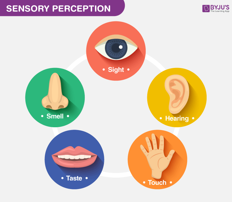
Five Sense Organs
As stated before, we have five sense organs that can receive and relay sensory information to the brain. These senses provide an organism with information crucial for perception. The different sense organs and the senses they provide are mentioned below:
Eyes – Sight or Ophthalmoception
These are the visual sensory organs in our body. These are sensitive to light images. The eyes vary in colour depending upon the amount of melanin present in our body. It helps in the sense of sight by detecting and focussing on the light images.
The iris in the eye is the coloured part that controls the size and diameter of the pupil, which directly affects the amount of light entering the eyes. Behind the lens of the eye lies the vitreous body. It is filled with a gelatinous material called the vitreous humour. This substance gives shape to the eyeball and also transmits light to the very back of the eyeball, where the retina is found.
This retina contains photoreceptors, which detect light. There are two types of cells present that perform functions distinct from each other. These are Rod and Cones.
Rods: These sensors function in low light and are found at the edges of the retina. They also aid in peripheral vision.
Cones: These types of retinal cells work best in bright light, detecting fine details and colour. There are three types of cones for detecting three primary colours of light, namely: blue, red and green. Typically, colour blindness occurs when any one of these types of cones are not present.
Also Read: Structure of the Eye
Ears – Hearing or Audioception
Ears are the auditory sense organs of our body. They help us to perceive sounds. Our auditory system detects vibrations in the air and this is how we hear sounds. This is known as hearing or audio caption.
The ears are divided into three sections, namely, the outer ear, the inner ear, and the middle ear. All sounds are basically vibrations, so the outer ear transfers these vibrations into the ear canal, where these vibrations are transformed by the brain into meaningful sound. Apart from hearing, this sense is also important for balancing our body or equilibrium.
Tongue – Taste or Gustaoception
The tongue helps in perceiving various tastes and flavours. The taste buds are present between the papillae on the tongue—these help in sensing different tastes.
The senses of smell and taste tend to work together. If one could not smell something, they could not taste it either. The sense of taste is also known as gustaoception.
Taste buds on the tongue contain chemoreceptors that work similarly to the chemoreceptors in the nasal cavity.
However, the chemoreceptors in the nose would detect any kind of smell, whereas there are four different types of taste buds and each one can detect different types of tastes like sweetness, sourness, bitterness and saltiness.
Discover: Interesting facts about tongue
Nose – Smell or Olfalcoception
The nose is an olfactory organ. Our olfactory system helps us to perceive different smells. This sense of organ also aids our sense of taste. The sense of smell is also known as olfaction.
The olfactory cells tend to line the top of the nasal cavity. On one end, olfactory cells have cilia that project into the nasal cavity and on the other end of the cell, are the olfactory nerve fibres.
As one breathes in, the air enters into the nasal cavity. The olfactory cells are chemoreceptors, which means that the olfactory cells have protein receptors that can detect subtle differences in chemicals. These chemicals bind to the cilia, which conducts a nerve impulse that is carried to the brain. The brain then translates these impulses into a meaningful smell. During a cold, the body produces mucus which blocks the sense of smell; this is the reason why the food which we eat tastes bland.
Skin – Touch or Tactioception
Skin is the largest organ of our body. It is related to the sense of touch. The sense of touch is also referred to as tactioception.
The skin contains general receptors which can detect touch, pain, pressure and temperature. They are present throughout the skin. Skin receptors generate an impulse, and when activated, is carried to the spinal cord and then to the brain.
Explore the Structure And Functions Of Skin
Other Sense Organs
Besides these five sense organs, there are another two that help to orient us with the world. They are:
Vestibular System
The vestibular system acts as a sensory system of the body and is responsible for transmitting information to our brain about the motions, head position and spatial orientation. This system is also involved with motor functions and helps in:
- Maintain our body posture.
- Maintaining our body balance.
- Stabilize our head and body during movement.
- Identifying the orientation and posture of our bodies in relation to the environment.
Thus, the vestibular system is essential for normal movement and equilibrium.
Proprioception system is described as the conscious or unconscious awareness of joint position. This system helps the body to identify the muscles, joints and limbs located in 3D space and the direction it is moving in relation to the body.
Walking or kicking without looking at our feet, balancing on one leg, touching the nose with eyes closed and the ability to sense the surface on which we are standing upon, are a few examples of proprioception system.
Further Reading: Sensory Perception
Learn more about sense organs and other related topics at BYJU’S Biology.
Students can register with BYJU’S Biology and access class-specific content such as sense organs chart, names of five sense organs, important questions and much more.
Frequently Asked Questions on Sense Organs
Define deafness., what is olfaction, what are the olfactory organs, which part of the human ear is responsible for maintaining body balance.

Put your understanding of this concept to test by answering a few MCQs. Click ‘Start Quiz’ to begin!
Select the correct answer and click on the “Finish” button Check your score and answers at the end of the quiz
Visit BYJU’S for all Biology related queries and study materials
Your result is as below
Request OTP on Voice Call
Leave a Comment Cancel reply
Your Mobile number and Email id will not be published. Required fields are marked *
Post My Comment
Thanks a lot, it is great
that was nice
Thank You Very Much For These Wonderful Experience.
Thank you a lot
Thanks, it’s good to study
Register with BYJU'S & Download Free PDFs
Register with byju's & watch live videos.
You are using an out of date browser. It may not display this or other websites correctly. You should upgrade or use an alternative browser .
Higher Order Questions for the Five Senses
Discussion in ' Kindergarten ' started by Sheila , Oct 28, 2008 .
Sheila Comrade
Oct 28, 2008
I have been trying to think of higher order-open ended questions in regards to the five senses. I am doing my observation on the five senses and have hit a rut! I am only able to come up with literal questions. Help!
Advertisement
janney Cohort
Which of the five senses is the most important? Why? Why do you think it is important that we be able to feel? Why do you think it is important that we be able to taste? Would you have a favorite food if you couldn't taste?
Share This Page
Members online now.

Receptors and Sense Organs
A sensory receptor is a neuron that detects stimuli. There are many kinds of sensory receptors. These receptors can be categorized based on the type of stimuli to which they respond.
• Mechanoreceptors respond to movement, pressure, and tension.
• Photoreceptors respond to variations in light.
• Chemoreceptors respond to chemicals.
• Thermoreceptors respond to changes in temperature.
• Pain receptors respond to tissue damage.
Sensory receptors are found in higher concentrations in the sense organs than in other parts of the body. When the sensory receptors of a particular sense organ receive appropriate stimulation, they convert the stimulus into electrical signals, or action potentials. These electrical signals are sent to specific regions of the brain. The action potentials generated by the different sense organs are electrically similar. So, how can a person know if the stimulation is a blue sky or a loud noise? The regions of the brain where the action potentials are interpreted vary according to the type of stimulus.
The brain has a specific region for each sense. Thus, signals received by the vision region of the occipital lobe are interpreted by the brain as images, even if the actual stimulus was something else. For example, a blow to the eye makes a person "see stars." The pressure of the blow stimulates visual neurons. The brain interprets this pressure as an image.
Continue reading here: Formation Of Sperm
Was this article helpful?
Related Posts
- Vaccines - Critical Thinking
- Meiosis In Grasshopper Testis
- The Grasshopper - Critical Thinking
- Collette Baughman Biology Teacher Benton Ks
- Protists In Industry - Critical Thinking
- Structure And Function Of Echinoderms
Readers' Questions
What is the difference between a sensory receptor and a sense organ?
A sensory receptor is a specialized cell or group of cells that respond to a specific stimulus, such as light, sound, heat, or pressure. Sense organs are collections of sensory receptors, such as the eyes, ears, nose, and tongue, which are responsible for collecting information from the outside environment and sending it to the brain.
What is the difference between sensory receptors and sense orgnas?
Sensory receptors are the cells or structures that receive and detect stimuli from the environment, while the senses are the organs that detect the stimulus from the environment and transfer them to the brain to be interpreted.
- Brain Development
- Childhood & Adolescence
- Diet & Lifestyle
- Emotions, Stress & Anxiety
- Learning & Memory
- Thinking & Awareness
- Alzheimer's & Dementia
- Childhood Disorders
- Immune System Disorders
- Mental Health
- Neurodegenerative Disorders
- Infectious Disease
- Neurological Disorders A-Z
- Body Systems
- Cells & Circuits
- Genes & Molecules
- The Arts & the Brain
- Law, Economics & Ethics
- Neuroscience in the News
- Supporting Research
- Tech & the Brain
- Animals in Research
- BRAIN Initiative
- Meet the Researcher
- Neuro-technologies
- Tools & Techniques
- Core Concepts
- For Educators
- Ask an Expert
- The Brain Facts Book
The Senses — A Primer (Part I)
- Published 11 Sep 2013
- Reviewed 11 Sep 2013
- Source The Dana Foundation
Our senses connect us to the world. Through complex systems that begin with cells that respond to physical stimuli and send signals through a maze of brain circuits, we can know—both consciously and otherwise—what goes on around us and within our bodies.
It’s a dynamic process. The brain is not simply a receiving station for sensory signals, and what we see, hear, and feel are constantly shaped by emotions, memories, moods, and beliefs. Our sense of the world is a creation of the brain, and the same physical sensation may be experienced quite differently at different times of life, and even from day to day.
The five (?) senses
We traditionally refer to the five senses of sight, hearing, taste, smell, and touch—a schema that dates back to Aristotle. But this is a simplification. We also have sensory systems to inform us of the position of our bodies (and parts of our bodies), visceral sensations, temperature, and pain, for example.
While each sensory system is unique, they share basic characteristics and similarities of structure and function. All are apparently active at birth, for example, although they may remain in a rudimentary state for weeks or months and continue to develop through childhood and adolescence. All are laid out along the same basic neural plan: a sense organ that turns physical phenomena like light, sound, or pressure into electrical impulses, and bundles of nerve fibers to carry these impulses to the brain. Sensory data generally pass through the thalamus, a kind of switching station atop the brain stem, en route to dedicated areas of the cortex designed to process them—the auditory cortex in the temporal lobe for hearing, for example, the visual cortex in the occipital lobe for sight. Smell—the oldest of the senses—is an exception: signals go directly from receptors in the nose to the olfactory bulb, in a more primitive part of the brain.
From these primary cortices, sensory information may engage wide and diverse areas of the brain: via direct connections with the limbic system, for example, an odor can trigger intense emotions; circuits that store memories give meaning to what we see and hear.
The senses have been studied extensively in an array of overlapping disciplines: physics and psychophysics, neuroanatomy, molecular biology, and cognitive psychology.
Sensory systems drew the attention of researchers early on, because their manifestations are more accessible to testing than most brain functions. The modern field of information science has become increasingly important to understanding how sensory processes extract and integrate multiple streams of data.
Stimulus and sensation
To equip humans for survival in their environment, the senses must be highly responsive—to signals as weak as a single photon of light or a molecule of an airborne chemical—and at the same time selective enough to filter information meriting attention from a noisy barrage of competing stimuli.
The process begins with anatomical features, such as the ear canals and the optics of the eye, designed to collect and channel stimuli to receptors that initiate the transduction process that turns them into electrical impulses. Typically, stimulation of a receptor cell (e.g. light on the retina) releases a protein that starts a biochemical cascade of messenger and energy-carrying molecules that generate electrical charges in a neuron, causing it to fire.
Receptors are highly specialized. In the retina, some cells (rods) respond to dim light, others (cones) to particular wavelengths; hair cells in the inner ear are tuned to different frequencies of sound. There are 350 subtypes of olfactory receptors, each responsive to a limited array of odors. Sense organs convey information about stimulus strength as well as quality: neurons fire faster as intensity increases.
From the resulting pattern of neural activity the brain derives details of sight, sound, smell, and other sensations.
From sensation to perception
Sensations themselves are fleeting: they linger in the memory for just seconds (sometimes less than a second) unless they engage neural networks beyond the primary sensory cortices. Perception is the process by which the brain makes sense of these incoming data, mixing memory, emotion, and cognition into the experience.
Many aspects of perception have been mapped out. Facial recognition is strongly associated with the fusiform face area in the temporal cortex, but areas of the occipital and prefrontal cortices, insula, and amygdala also take part in the complex process that distinguishes faces from other objects, identifies emotional expressions, and recognizes familiar individuals. The shapes of letters engage a nearby part of the visual system, the visual word form area.
Perception involves “top-down” as well as “bottom-up” processing. That is, higher brain areas don’t just respond to sensory information, they actively condition it: inhibiting irrelevant input, for example, and completing meaningful structures from fragments—e.g. constructing words from partial sounds. We can follow a conversation at a noisy party because our brains are doing more than simply translating auditory sensations. Indeed, there is evidence that “mindset”—expectation and attitude—can modify neuron firing in primary sensory cortices: we see and hear what we think we will.
Emotion likewise amplifies sensory processing at an early stage. One recent study found that scenes look particularly vivid—and activity rises in the visual cortex—at times of emotional arousal. Another study reported that anxious men could detect threatening odors at a lower concentration than the non-anxious, reflecting amped-up activity in primary olfactory centers.
Senses and neuroplasticity
Although the brain’s sensory systems are wired from before birth, they continually evolve through interaction with the environment.
The senses need stimulation during early “critical periods” to form necessary neural connections. If an eye is covered for the first six months of life, it may remain functionally blind even after it is uncovered. To an extent, adult neuroplasticity can mitigate the loss. People blind from birth whose sight was restored later in life can learn to make sense of visual input, although it is a painstaking and imperfect process.
Sensory training makes a difference in more subtle ways. While “perfect pitch” probably requires genetic endowment, early sound exposure apparently plays a role as well—many more children develop this capacity in Asian countries where pitch is an essential element of language than in the West. Even in adulthood, the ability to discriminate pitch improves with practice.
Visual skills can be similarly sharpened. A recent study suggested that artistic training doesn’t alter activity in the visual cortex itself, but refines the ability of higher brain areas to process this information.
A most striking demonstration of sensory neuroplasticity is cross-modal compensation: the loss of one sense heightens another. The enhanced hearing and touch of many sight-impaired people reflects the reassignment of parts of the visual cortex to auditory and tactile processing, according to neuroimaging and electrophysiological studies. Reportedly, some blind individuals even learn to use “echolocation” to navigate their way around objects by listening to reflected sounds, using repurposed brain areas that ordinarily process sight.
- By Carl Sherman
CONTENT PROVIDED BY
The Dana Foundation
The Dana Foundation is a private philanthropic organization that supports brain research through grants and educates the public about the successes and potential of brain research.
Stein, J.F. Neuroscience: an introduction. Wiley. 2006 Henshaw, J. M. A Tour of the Senses: how your brain interprets the world. Johns Hopkins University Press. 2012 Bossomaier, T. R. J. Introduction to the Senses: from biology to computer science. Cambridge University Press. 2012 Brynie, F.H. Brain Sense: the science of the senses and how we process the world around us. Amacom. 2009 The Brain from Top to Bottom: The Senses: Vision. Canadian Institute of Health Research; Institute of Neurosciences, Mental Health and Addiction. http://thebrain.mcgill.ca/flash/a/a_02/a_02_cr/a_02_cr_vis/a_02_cr_vis.html Lu, Z.-L., & Sperling, G. Measuring sensory memory: Magnetoencephalography habituation and psychophysics. In Z.-L. Lu & L. Kaufman (Eds.) Magnetic source imaging of the human brain. Mahwah, NJ: Lawrence Erlbaum Associates, Inc. 2003. Pp. 319-342. http://lobes.usc.edu/Journals/LEA03b.pdf Gobbini MI, Haxby JV. . Neural systems for recognition of familiar faces. Neuropsychologia. 2007 Jan 7;45(1):32-41. Epub 2006 Jun 22 Pyles JA, Verstynen TD, et al. Explicating the face perception network with white matter connectivity. PLoS One. 2013 Apr 22;8(4):e61611. Dehaene, S. Inside the Letterbox: How Literacy Transforms the Human Brain. Cerebrum. June 03, 2013. Krusemark EA, Li W. Enhanced Olfactory Sensory Perception of Threat in Anxiety: An Event-Related fMRI Study. Chemosens Percept. 2012 Mar 1;5(1):37-45. Epub 2012 Jan 10. Todd, R. M., Talmi, D., et al. Psychophysical and Neural Evidence for Emotion-Enhanced Perceptual Vividness. J Neuroscience, 15 August 2012, 32(33): 11201-11212. Constantine-Paton M. Pioneers of cortical plasticity: six classic papers by Wiesel and Hubel. J Neurophysiol. 2008 Jun;99(6):2741-4. Zatorre, RA, Absolute pitch: a model for understanding the influence of genes and development on neural and cognitive function. Nature Neuroscience (July 2003); 6(7): 692-695. Perdreau, F, & Cavanagh, P. Is artists’ perception more veridical? Frontiers in Neurosci. (January 2013). 7:1-11. Pascual-Leone, Amedi, A et al , The Plastic Human Brain Cortex. Annu. Rev. Neurosci. 2005. 28:377–401. Thaler, L, Arnott, S. R., Goodale, M. A. Neural Correlates of Natural Human Echolocation in Early and Late Blind Echolocation Experts. PLoS ONE May 2011; 6 (5): e20162.
Also In Vision

Popular articles on BrainFacts.org

BrainFacts Book
Download a copy of the newest edition of the book, Brain Facts: A Primer on the Brain and Nervous System.
Research & Discoveries
See how discoveries in the lab have improved human health.
Personalize Your Emails
Personalize your monthly updates from BrainFacts.org by choosing the topics that you care about most!

SUPPORTING PARTNERS
- Accessibility Policy
- Terms and Conditions
- Manage Cookies
Some pages on this website provide links that require Adobe Reader to view.

Environment
The five senses and the nature of perception, perceiving the world looks, sounds, and feels easy. it isn't..
Posted November 11, 2014 | Reviewed by Jessica Schrader
We perceive the world through our five senses—our eyes, ears, skin, nose, and mouth are all receptors. Everything that comes into the brain enters through one of these doors. Because most of us take the world in through our senses effortlessly, we don’t give much thought or attention to how we do this.
Even scientists were guilty of underappreciating the complexity of the senses. Back in the 1950s and 1960s, when computers were in their infancy, the thinking was that it would take a decade or so to build “perceiving machines” that could respond to sight, sound, touch and so on as well as a human being. Such a machine still doesn’t exist.
Lose a sense, however, and you will quickly appreciate what is missing. I know because that’s what happened to me when I found out my son was deaf. There was so much to learn about the way hearing works and the role of sound in the brain that I wrote a whole book about it. That was the long version.
This is the short version. What has to happen to put on the show that is our awareness of our environment? An awful lot. Neuroscientists have recently done some radical rethinking about the very nature of perception .
“Historically, the way we intuitively think about all perception is that we’re like a passive recording device with detectors that are specialized for certain things, like a retina for seeing, a cochlea for hearing, and so forth,” says David Poeppel , a professor of psychology and neural science at New York University and a director of the newly established Max Planck Institute for Empirical Aesthetics . “We’re kind of a camera or microphone that gets encoded somehow and then magically makes contact with the stuff in your head.”
At the same time, many of the big thinkers who pondered perception, as far back as the 19th-century German physician Hermann von Helmholtz, knew that couldn’t be quite right. If we reached for a glass or listened to a sentence, didn’t it help to be able to anticipate what might come next?
In the mid-to-late 20th century, a handful of prominent researchers proposed models of perception that suggested that we engaged in “active sensing,” seeking out what was possible as we went along. Such ideas didn’t gain much traction until the past decade, when they suddenly became a hot topic in the study of cognition . What everyone is talking about today is the brain’s power of prediction.
On one level, prediction is just common sense, which may be one reason it didn’t get much scientific respect for so long. If you see your doctor in the doctor’s office, you recognize her quickly. If you see her in the grocery store dressed in jeans, you’ll be slower to realize you know her.
Predictable events are easy for the brain; unpredictable events require more effort. “Our expectations for what we’re going to perceive seem to be a critical part of the process,” says Greg Hickok , a neuroscientist at the University of California, Irvine. “It allows the system to make guesses as to what it might be seeing and to use computational shortcuts.”
In the old view of perception, a cascade of responses flows from the ear or the eye through the brain and ends with the ability to follow a complicated sentence or pick out the one person you are looking for in a crowded theater. That is known as bottom-up processing. It starts with basic input to any sense—raw data—and ends with such higher-level skills as reasoning and judgment and critical thinking—in other words, our expectations and knowledge.
But that is only half the story. Neuroscientists now believe that the process is also happening in reverse, that the cascade flows both ways, with information being prepared, treated, and converted in both directions simultaneously, from the bottom up and the top down.
This holds for simple responses as well as for complex thinking about philosophy or physics. If a sound is uncomfortably loud, for instance, it is the cortex that registers that fact and sends a message all the way back to the cochlea to stiffen hair cells as a protective measure. The same is true of the retina, adjusting for the amount of light available. It’s not your eye or ear doing that, it’s your brain.
Imagine someone beating rhythmically on a table with a pencil: tap, tap, tap, tap. By the third beat, you have anticipated the timing. By the fourth, scientists like Poeppel and Hickok could see activity in the brain that represents that prediction.
Perception then is an active process of constructing a reality, a conversation between the senses and the cortex that balances new information from the outside world with predictions from the interior world of our brain.

Parts of this post originally appeared in I Can Hear You Whisper: An Intimate Journey through the Science of Sound and Language (Dutton 2014).

Lydia Denworth is a science journalist and author of Friendship: The Evolution, Biology, and Extraordinary Power of Life’s Fundamental Bond.
- Find a Therapist
- Find a Treatment Center
- Find a Psychiatrist
- Find a Support Group
- Find Online Therapy
- United States
- Brooklyn, NY
- Chicago, IL
- Houston, TX
- Los Angeles, CA
- New York, NY
- Portland, OR
- San Diego, CA
- San Francisco, CA
- Seattle, WA
- Washington, DC
- Asperger's
- Bipolar Disorder
- Chronic Pain
- Eating Disorders
- Passive Aggression
- Personality
- Goal Setting
- Positive Psychology
- Stopping Smoking
- Low Sexual Desire
- Relationships
- Child Development
- Self Tests NEW
- Therapy Center
- Diagnosis Dictionary
- Types of Therapy

It’s increasingly common for someone to be diagnosed with a condition such as ADHD or autism as an adult. A diagnosis often brings relief, but it can also come with as many questions as answers.
- Emotional Intelligence
- Gaslighting
- Affective Forecasting
- Neuroscience
13.2 The Central Nervous System
Learning objectives.
By the end of this section, you will be able to:
- Name the major regions of the adult brain
- Describe the connections between the cerebrum and brain stem through the diencephalon, and from those regions into the spinal cord
- Recognize the complex connections within the subcortical structures of the basal nuclei
- Explain the arrangement of gray and white matter in the spinal cord
The brain and the spinal cord are the central nervous system, and they represent the main organs of the nervous system. The spinal cord is a single structure, whereas the adult brain is described in terms of four major regions: the cerebrum, the diencephalon, the brain stem, and the cerebellum. A person’s conscious experiences are based on neural activity in the brain. The regulation of homeostasis is governed by a specialized region in the brain. The coordination of reflexes depends on the integration of sensory and motor pathways in the spinal cord.
The Cerebrum
The iconic gray mantle of the human brain, which appears to make up most of the mass of the brain, is the cerebrum ( Figure 13.6 ). The wrinkled portion is the cerebral cortex , and the rest of the structure is beneath that outer covering. There is a large separation between the two sides of the cerebrum called the longitudinal fissure . It separates the cerebrum into two distinct halves, a right and left cerebral hemisphere . Deep within the cerebrum, the white matter of the corpus callosum provides the major pathway for communication between the two hemispheres of the cerebral cortex.
Many of the higher neurological functions, such as memory, emotion, and consciousness, are the result of cerebral function. The complexity of the cerebrum is different across vertebrate species. The cerebrum of the most primitive vertebrates is not much more than the connection for the sense of smell. In mammals, the cerebrum comprises the outer gray matter that is the cortex (from the Latin word meaning “bark of a tree”) and several deep nuclei that belong to three important functional groups. The basal nuclei are responsible for cognitive processing, the most important function being that associated with planning movements. The basal forebrain contains nuclei that are important in learning and memory. The limbic cortex is the region of the cerebral cortex that is part of the limbic system , a collection of structures involved in emotion, memory, and behavior.
Cerebral Cortex
The cerebrum is covered by a continuous layer of gray matter that wraps around either side of the forebrain—the cerebral cortex. This thin, extensive region of wrinkled gray matter is responsible for the higher functions of the nervous system. A gyrus (plural = gyri) is the ridge of one of those wrinkles, and a sulcus (plural = sulci) is the groove between two gyri. The pattern of these folds of tissue indicates specific regions of the cerebral cortex.
The head is limited by the size of the birth canal, and the brain must fit inside the cranial cavity of the skull. Extensive folding in the cerebral cortex enables more gray matter to fit into this limited space. If the gray matter of the cortex were peeled off of the cerebrum and laid out flat, its surface area would be roughly equal to one square meter.
The folding of the cortex maximizes the amount of gray matter in the cranial cavity. During embryonic development, as the telencephalon expands within the skull, the brain goes through a regular course of growth that results in everyone’s brain having a similar pattern of folds. The surface of the brain can be mapped on the basis of the locations of large gyri and sulci. Using these landmarks, the cortex can be separated into four major regions, or lobes ( Figure 13.7 ). The lateral sulcus that separates the temporal lobe from the other regions is one such landmark. Superior to the lateral sulcus are the parietal lobe and frontal lobe , which are separated from each other by the central sulcus . The posterior region of the cortex is the occipital lobe , which has no obvious anatomical border between it and the parietal or temporal lobes on the lateral surface of the brain. From the medial surface, an obvious landmark separating the parietal and occipital lobes is called the parieto-occipital sulcus . The fact that there is no obvious anatomical border between these lobes is consistent with the functions of these regions being interrelated.
Different regions of the cerebral cortex can be associated with particular functions, a concept known as localization of function. In the early 1900s, a German neuroscientist named Korbinian Brodmann performed an extensive study of the microscopic anatomy—the cytoarchitecture—of the cerebral cortex and divided the cortex into 52 separate regions on the basis of the histology of the cortex. His work resulted in a system of classification known as Brodmann’s areas , which is still used today to describe the anatomical distinctions within the cortex ( Figure 13.8 ). The results from Brodmann’s work on the anatomy align very well with the functional differences within the cortex. Areas 17 and 18 in the occipital lobe are responsible for primary visual perception. That visual information is complex, so it is processed in the temporal and parietal lobes as well.
The temporal lobe is associated with primary auditory sensation, known as Brodmann’s areas 41 and 42 in the superior temporal lobe. Because regions of the temporal lobe are part of the limbic system, memory is an important function associated with that lobe. Memory is essentially a sensory function; memories are recalled sensations such as the smell of Mom’s baking or the sound of a barking dog. Even memories of movement are really the memory of sensory feedback from those movements, such as stretching muscles or the movement of the skin around a joint. Structures in the temporal lobe are responsible for establishing long-term memory, but the ultimate location of those memories is usually in the region in which the sensory perception was processed.
The main sensation associated with the parietal lobe is somatosensation , meaning the general sensations associated with the body. Posterior to the central sulcus is the postcentral gyrus , the primary somatosensory cortex, which is identified as Brodmann’s areas 1, 2, and 3. All of the tactile senses are processed in this area, including touch, pressure, tickle, pain, itch, and vibration, as well as more general senses of the body such as proprioception and kinesthesia , which are the senses of body position and movement, respectively.
Anterior to the central sulcus is the frontal lobe, which is primarily associated with motor functions. The precentral gyrus is the primary motor cortex. Cells from this region of the cerebral cortex are the upper motor neurons that instruct cells in the spinal cord to move skeletal muscles. Anterior to this region are a few areas that are associated with planned movements. The premotor area is responsible for thinking of a movement to be made. The frontal eye fields are important in eliciting eye movements and in attending to visual stimuli. Broca’s area is responsible for the production of language, or controlling movements responsible for speech; in the vast majority of people, it is located only on the left side. Anterior to these regions is the prefrontal lobe , which serves cognitive functions that can be the basis of personality, short-term memory, and consciousness. The prefrontal lobotomy is an outdated mode of treatment for personality disorders (psychiatric conditions) that profoundly affected the personality of the patient.
Subcortical structures
Beneath the cerebral cortex are sets of nuclei known as subcortical nuclei that augment cortical processes. The nuclei of the basal forebrain serve as the primary location for acetylcholine production, which modulates the overall activity of the cortex, possibly leading to greater attention to sensory stimuli. Alzheimer’s disease is associated with a loss of neurons in the basal forebrain. The hippocampus and amygdala are medial-lobe structures that, along with the adjacent cortex, are involved in long-term memory formation and emotional responses. The basal nuclei are a set of nuclei in the cerebrum responsible for comparing cortical processing with the general state of activity in the nervous system to influence the likelihood of movement taking place. For example, while a student is sitting in a classroom listening to a lecture, the basal nuclei will keep the urge to jump up and scream from actually happening. (The basal nuclei are also referred to as the basal ganglia, although that is potentially confusing because the term ganglia is typically used for peripheral structures.)
The major structures of the basal nuclei that control movement are the caudate , putamen , and globus pallidus , which are located deep in the cerebrum. The caudate is a long nucleus that follows the basic C-shape of the cerebrum from the frontal lobe, through the parietal and occipital lobes, into the temporal lobe. The putamen is mostly deep in the anterior regions of the frontal and parietal lobes. Together, the caudate and putamen are called the striatum . The globus pallidus is a layered nucleus that lies just medial to the putamen; they are called the lenticular nuclei because they look like curved pieces fitting together like lenses. The globus pallidus has two subdivisions, the external and internal segments, which are lateral and medial, respectively. These nuclei are depicted in a frontal section of the brain in Figure 13.9 .
The basal nuclei in the cerebrum are connected with a few more nuclei in the brain stem that together act as a functional group that forms a motor pathway. Two streams of information processing take place in the basal nuclei. All input to the basal nuclei is from the cortex into the striatum ( Figure 13.10 ). The direct pathway is the projection of axons from the striatum to the globus pallidus internal segment (GPi) and the substantia nigra pars reticulata (SNr). The GPi/SNr then projects to the thalamus, which projects back to the cortex. The indirect pathway is the projection of axons from the striatum to the globus pallidus external segment (GPe), then to the subthalamic nucleus (STN), and finally to GPi/SNr. The two streams both target the GPi/SNr, but one has a direct projection and the other goes through a few intervening nuclei. The direct pathway causes the disinhibition of the thalamus (inhibition of one cell on a target cell that then inhibits the first cell), whereas the indirect pathway causes, or reinforces, the normal inhibition of the thalamus. The thalamus then can either excite the cortex (as a result of the direct pathway) or fail to excite the cortex (as a result of the indirect pathway).
The switch between the two pathways is the substantia nigra pars compacta , which projects to the striatum and releases the neurotransmitter dopamine. Dopamine receptors are either excitatory (D1-type receptors) or inhibitory (D2-type receptors). The direct pathway is activated by dopamine, and the indirect pathway is inhibited by dopamine. When the substantia nigra pars compacta is firing, it signals to the basal nuclei that the body is in an active state, and movement will be more likely. When the substantia nigra pars compacta is silent, the body is in a passive state, and movement is inhibited. To illustrate this situation, while a student is sitting listening to a lecture, the substantia nigra pars compacta would be silent and the student less likely to get up and walk around. Likewise, while the professor is lecturing, and walking around at the front of the classroom, the professor’s substantia nigra pars compacta would be active, in keeping with their activity level.
Interactive Link
Watch this video to learn about the basal nuclei (also known as the basal ganglia), which have two pathways that process information within the cerebrum. As shown in this video, the direct pathway is the shorter pathway through the system that results in increased activity in the cerebral cortex and increased motor activity. The direct pathway is described as resulting in “disinhibition” of the thalamus. What does disinhibition mean? What are the two neurons doing individually to cause this?
Watch this video to learn about the basal nuclei (also known as the basal ganglia), which have two pathways that process information within the cerebrum. As shown in this video, the indirect pathway is the longer pathway through the system that results in decreased activity in the cerebral cortex, and therefore less motor activity. The indirect pathway has an extra couple of connections in it, including disinhibition of the subthalamic nucleus. What is the end result on the thalamus, and therefore on movement initiated by the cerebral cortex?
Everyday Connection
The myth of left brain/right brain.
There is a persistent myth that people are “right-brained” or “left-brained,” which is an oversimplification of an important concept about the cerebral hemispheres. There is some lateralization of function, in which the left side of the brain is devoted to language function and the right side is devoted to spatial and nonverbal reasoning. Whereas these functions are predominantly associated with those sides of the brain, there is no monopoly by either side on these functions. Many pervasive functions, such as language, are distributed globally around the cerebrum.
Some of the support for this misconception has come from studies of split brains. A drastic way to deal with a rare and devastating neurological condition (intractable epilepsy) is to separate the two hemispheres of the brain. After sectioning the corpus callosum, a split-brained patient will have trouble producing verbal responses on the basis of sensory information processed on the right side of the cerebrum, leading to the idea that the left side is responsible for language function.
However, there are well-documented cases of language functions lost from damage to the right side of the brain. The deficits seen in damage to the left side of the brain are classified as aphasia, a loss of speech function; damage on the right side can affect the use of language. Right-side damage can result in a loss of ability to understand figurative aspects of speech, such as jokes, irony, or metaphors. Nonverbal aspects of speech can be affected by damage to the right side, such as facial expression or body language, and right-side damage can lead to a “flat affect” in speech, or a loss of emotional expression in speech—sounding like a robot when talking.
The Diencephalon
The diencephalon is the one region of the adult brain that retains its name from embryologic development. The etymology of the word diencephalon translates to “through brain.” It is the connection between the cerebrum and the rest of the nervous system, with one exception. The rest of the brain, the spinal cord, and the PNS all send information to the cerebrum through the diencephalon. Output from the cerebrum passes through the diencephalon. The single exception is the system associated with olfaction , or the sense of smell, which connects directly with the cerebrum. In the earliest vertebrate species, the cerebrum was not much more than olfactory bulbs that received peripheral information about the chemical environment (to call it smell in these organisms is imprecise because they lived in the ocean).
The diencephalon is deep beneath the cerebrum and constitutes the walls of the third ventricle. The diencephalon can be described as any region of the brain with “thalamus” in its name. The two major regions of the diencephalon are the thalamus itself and the hypothalamus ( Figure 13.11 ). There are other structures, such as the epithalamus , which contains the pineal gland, or the subthalamus , which includes the subthalamic nucleus that is part of the basal nuclei.
The thalamus is a collection of nuclei that relay information between the cerebral cortex and the periphery, spinal cord, or brain stem. All sensory information, except for the sense of smell, passes through the thalamus before processing by the cortex. Axons from the peripheral sensory organs, or intermediate nuclei, synapse in the thalamus, and thalamic neurons project directly to the cerebrum. It is a requisite synapse in any sensory pathway, except for olfaction. The thalamus does not just pass the information on, it also processes that information. For example, the portion of the thalamus that receives visual information will influence what visual stimuli are important, or what receives attention.
The cerebrum also sends information down to the thalamus, which usually communicates motor commands. This involves interactions with the cerebellum and other nuclei in the brain stem. The cerebrum interacts with the basal nuclei, which involves connections with the thalamus. The primary output of the basal nuclei is to the thalamus, which relays that output to the cerebral cortex. The cortex also sends information to the thalamus that will then influence the effects of the basal nuclei.
Hypothalamus
Inferior and slightly anterior to the thalamus is the hypothalamus , the other major region of the diencephalon. The hypothalamus is a collection of nuclei that are largely involved in regulating homeostasis. The hypothalamus is the executive region in charge of the autonomic nervous system and the endocrine system through its regulation of the anterior pituitary gland. Other parts of the hypothalamus are involved in memory and emotion as part of the limbic system.
The midbrain and hindbrain (composed of the pons and the medulla) are collectively referred to as the brain stem ( Figure 13.12 ). The structure emerges from the ventral surface of the forebrain as a tapering cone that connects the brain to the spinal cord. Attached to the brain stem, but considered a separate region of the adult brain, is the cerebellum. The midbrain coordinates sensory representations of the visual, auditory, and somatosensory perceptual spaces. The pons is the main connection with the cerebellum. The pons and the medulla regulate several crucial functions, including the cardiovascular and respiratory systems and rates.
The cranial nerves connect through the brain stem and provide the brain with the sensory input and motor output associated with the head and neck, including most of the special senses. The major ascending and descending pathways between the spinal cord and brain, specifically the cerebrum, pass through the brain stem.
One of the original regions of the embryonic brain, the midbrain is a small region between the thalamus and pons. It is separated into the tectum and tegmentum , from the Latin words for roof and floor, respectively. The cerebral aqueduct passes through the center of the midbrain, such that these regions are the roof and floor of that canal.
The tectum is composed of four bumps known as the colliculi (singular = colliculus), which means “little hill” in Latin. The inferior colliculus is the inferior pair of these enlargements and is part of the auditory brain stem pathway. Neurons of the inferior colliculus project to the thalamus, which then sends auditory information to the cerebrum for the conscious perception of sound. The superior colliculus is the superior pair and combines sensory information about visual space, auditory space, and somatosensory space. Activity in the superior colliculus is related to orienting the eyes to a sound or touch stimulus. If you are walking along the sidewalk on campus and you hear chirping, the superior colliculus coordinates that information with your awareness of the visual location of the tree right above you. That is the correlation of auditory and visual maps. If you suddenly feel something wet fall on your head, your superior colliculus integrates that with the auditory and visual maps and you know that the chirping bird just relieved itself on you. You want to look up to see the culprit, but do not.
The tegmentum is continuous with the gray matter of the rest of the brain stem. Throughout the midbrain, pons, and medulla, the tegmentum contains the nuclei that receive and send information through the cranial nerves, as well as regions that regulate important functions such as those of the cardiovascular and respiratory systems.
The word pons comes from the Latin word for bridge. It is visible on the anterior surface of the brain stem as the thick bundle of white matter attached to the cerebellum. The pons is the main connection between the cerebellum and the brain stem. The bridge-like white matter is only the anterior surface of the pons; the gray matter beneath that is a continuation of the tegmentum from the midbrain. Gray matter in the tegmentum region of the pons contains neurons receiving descending input from the forebrain that is sent to the cerebellum.
The medulla is the region known as the myelencephalon in the embryonic brain. The initial portion of the name, “myel,” refers to the significant white matter found in this region—especially on its exterior, which is continuous with the white matter of the spinal cord. The tegmentum of the midbrain and pons continues into the medulla because this gray matter is responsible for processing cranial nerve information. A diffuse region of gray matter throughout the brain stem, known as the reticular formation , is related to sleep and wakefulness, such as general brain activity and attention.
The Cerebellum
The cerebellum , as the name suggests, is the “little brain.” It is covered in gyri and sulci like the cerebrum, and looks like a miniature version of that part of the brain ( Figure 13.13 ). The cerebellum is largely responsible for comparing information from the cerebrum with sensory feedback from the periphery through the spinal cord. It accounts for approximately 10 percent of the mass of the brain.
Descending fibers from the cerebrum have branches that connect to neurons in the pons. Those neurons project into the cerebellum, providing a copy of motor commands sent to the spinal cord. Sensory information from the periphery, which enters through spinal or cranial nerves, is copied to a nucleus in the medulla known as the inferior olive . Fibers from this nucleus enter the cerebellum and are compared with the descending commands from the cerebrum. If the primary motor cortex of the frontal lobe sends a command down to the spinal cord to initiate walking, a copy of that instruction is sent to the cerebellum. Sensory feedback from the muscles and joints, proprioceptive information about the movements of walking, and sensations of balance are sent to the cerebellum through the inferior olive and the cerebellum compares them. If walking is not coordinated, perhaps because the ground is uneven or a strong wind is blowing, then the cerebellum sends out a corrective command to compensate for the difference between the original cortical command and the sensory feedback. The output of the cerebellum is into the midbrain, which then sends a descending input to the spinal cord to correct the messages going to skeletal muscles.
The Spinal Cord
The description of the CNS is concentrated on the structures of the brain, but the spinal cord is another major organ of the system. Whereas the brain develops out of expansions of the neural tube into primary and then secondary vesicles, the spinal cord maintains the tube structure and is only specialized into certain regions. As the spinal cord continues to develop in the newborn, anatomical features mark its surface. The anterior midline is marked by the anterior median fissure , and the posterior midline is marked by the posterior median sulcus . Axons enter the posterior side through the dorsal (posterior) nerve root , which marks the posterolateral sulcus on either side. The axons emerging from the anterior side do so through the ventral (anterior) nerve root . Note that it is common to see the terms dorsal (dorsal = “back”) and ventral (ventral = “belly”) used interchangeably with posterior and anterior, particularly in reference to nerves and the structures of the spinal cord. You should learn to be comfortable with both.
On the whole, the posterior regions are responsible for sensory functions and the anterior regions are associated with motor functions. This comes from the initial development of the spinal cord, which is divided into the basal plate and the alar plate . The basal plate is closest to the ventral midline of the neural tube, which will become the anterior face of the spinal cord and gives rise to motor neurons. The alar plate is on the dorsal side of the neural tube and gives rise to neurons that will receive sensory input from the periphery.
The length of the spinal cord is divided into regions that correspond to the regions of the vertebral column. The name of a spinal cord region corresponds to the level at which spinal nerves pass through the intervertebral foramina. Immediately adjacent to the brain stem is the cervical region, followed by the thoracic, then the lumbar, and finally the sacral region. The spinal cord is not the full length of the vertebral column because the spinal cord does not grow significantly longer after the first or second year, but the skeleton continues to grow. The nerves that emerge from the spinal cord pass through the intervertebral foramina at the respective levels. As the vertebral column grows, these nerves grow with it and result in a long bundle of nerves that resembles a horse’s tail and is named the cauda equina . The sacral spinal cord is at the level of the upper lumbar vertebral bones. The spinal nerves extend from their various levels to the proper level of the vertebral column.
In cross-section, the gray matter of the spinal cord has the appearance of an ink-blot test, with the spread of the gray matter on one side replicated on the other—a shape reminiscent of a bulbous capital “H.” As shown in Figure 13.14 , the gray matter is subdivided into regions that are referred to as horns. The posterior horn is responsible for sensory processing. The anterior horn sends out motor signals to the skeletal muscles. The lateral horn , which is only found in the thoracic, upper lumbar, and sacral regions, contains cell bodies of motor neurons of the autonomic nervous system.
Some of the largest neurons of the spinal cord are the multipolar motor neurons in the anterior horn. The fibers that cause contraction of skeletal muscles are the axons of these neurons. The motor neuron that causes contraction of the big toe, for example, is located in the sacral spinal cord. The axon that has to reach all the way to the belly of that muscle may be a meter in length. The neuronal cell body that maintains that long fiber must be quite large, possibly several hundred micrometers in diameter, making it one of the largest cells in the body.
White Columns
Just as the gray matter is separated into horns, the white matter of the spinal cord is separated into columns. Ascending tracts of nervous system fibers in these columns carry sensory information up to the brain, whereas descending tracts carry motor commands from the brain. Looking at the spinal cord longitudinally, the columns extend along its length as continuous bands of white matter. Between the two posterior horns of gray matter are the posterior columns . Between the two anterior horns, and bounded by the axons of motor neurons emerging from that gray matter area, are the anterior columns . The white matter on either side of the spinal cord, between the posterior horn and the axons of the anterior horn neurons, are the lateral columns . The posterior columns are composed of axons of ascending tracts. The anterior and lateral columns are composed of many different groups of axons of both ascending and descending tracts—the latter carrying motor commands down from the brain to the spinal cord to control output to the periphery.
Watch this video to learn about the gray matter of the spinal cord that receives input from fibers of the dorsal (posterior) root and sends information out through the fibers of the ventral (anterior) root. As discussed in this video, these connections represent the interactions of the CNS with peripheral structures for both sensory and motor functions. The cervical and lumbar spinal cords have enlargements as a result of larger populations of neurons. What are these enlargements responsible for?
Disorders of the...
Basal nuclei.
Parkinson’s disease is a disorder of the basal nuclei, specifically of the substantia nigra, that demonstrates the effects of the direct and indirect pathways. Parkinson’s disease is the result of neurons in the substantia nigra pars compacta dying. These neurons release dopamine into the striatum. Without that modulatory influence, the basal nuclei are stuck in the indirect pathway, without the direct pathway being activated. The direct pathway is responsible for increasing cortical movement commands. The increased activity of the indirect pathway results in the hypokinetic disorder of Parkinson’s disease.
Parkinson’s disease is neurodegenerative, meaning that neurons die that cannot be replaced, so there is no cure for the disorder. Treatments for Parkinson’s disease are aimed at increasing dopamine levels in the striatum. Currently, the most common way of doing that is by providing the amino acid L-DOPA, which is a precursor to the neurotransmitter dopamine and can cross the blood-brain barrier. With levels of the precursor elevated, the remaining cells of the substantia nigra pars compacta can make more neurotransmitter and have a greater effect. Unfortunately, the patient will become less responsive to L-DOPA treatment as time progresses, and it can cause increased dopamine levels elsewhere in the brain, which are associated with psychosis or schizophrenia.
Visit this site for a thorough explanation of Parkinson’s disease.
Compared with the nearest evolutionary relative, the chimpanzee, the human has a brain that is huge. At a point in the past, a common ancestor gave rise to the two species of humans and chimpanzees. That evolutionary history is long and is still an area of intense study. But something happened to increase the size of the human brain relative to the chimpanzee. Read this article in which the author explores the current understanding of why this happened.
According to one hypothesis about the expansion of brain size, what tissue might have been sacrificed so energy was available to grow our larger brain? Based on what you know about that tissue and nervous tissue, why would there be a trade-off between them in terms of energy use?
This book may not be used in the training of large language models or otherwise be ingested into large language models or generative AI offerings without OpenStax's permission.
Want to cite, share, or modify this book? This book uses the Creative Commons Attribution License and you must attribute OpenStax.
Access for free at https://openstax.org/books/anatomy-and-physiology-2e/pages/1-introduction
- Authors: J. Gordon Betts, Kelly A. Young, James A. Wise, Eddie Johnson, Brandon Poe, Dean H. Kruse, Oksana Korol, Jody E. Johnson, Mark Womble, Peter DeSaix
- Publisher/website: OpenStax
- Book title: Anatomy and Physiology 2e
- Publication date: Apr 20, 2022
- Location: Houston, Texas
- Book URL: https://openstax.org/books/anatomy-and-physiology-2e/pages/1-introduction
- Section URL: https://openstax.org/books/anatomy-and-physiology-2e/pages/13-2-the-central-nervous-system
© Jun 13, 2024 OpenStax. Textbook content produced by OpenStax is licensed under a Creative Commons Attribution License . The OpenStax name, OpenStax logo, OpenStax book covers, OpenStax CNX name, and OpenStax CNX logo are not subject to the Creative Commons license and may not be reproduced without the prior and express written consent of Rice University.
IMAGES
VIDEO
COMMENTS
As we know, the sense organs are the organs in the body that respond to external stimuli by conveying impulses to the sensory nervous system. These organs are responsible for our perception of sounds, smell, sight, taste, and touch. Try this short and simple quiz today and see how much you remember about your organs. Questions and Answers. 1.
Study with Quizlet and memorize flashcards containing terms like name the 5 most familiar sense organs, how are sensory receptors classified, name the 5 groups of sensory receptors and more. ... Biology 1 - Chapter 14 Critical Thinking Questions. 13 terms. jkglaspey. Preview. ELA the Call of the Wild Vocab list. 30 terms. roykim090108. Preview ...
Of course, other materials may be substituted at each station.) Your five senses — seeing, hearing, smelling, tasting, and touching — help you notice the world around you. They're pretty powerful! You use your eyes to see, your ears to hear, your nose to smell, your tongue to taste, and your skin to feel.
the colored part of the eye. the lining at the back of the eyeball. sends messages to the brain. clear layer at the front-center of the eye. 45 s. Quiz your students on Grade 2 - The Five Sense Organs practice problems using our fun classroom quiz game Quizalize and personalize your teaching.
unit. See Section XI for text-based critical thinking questions to support the read aloud experience. Engaging, informative, and literary texts provide opportunities for exploring content, expressing ideas using one's imagination, and critical thinking that are enhanced through multiple readings of the same book.
Which plane divides the heart into anterior and posterior portions? parasagittal plane. Which plane divides the brain into unequal right and left portions? Urinary bladder = P, stomach = A, heart = T, small intestine = A, lungs = T, internal female reproductive organs = P, thymus = T, spleen = A, liver = A. In which cavities are the following ...
Introduction ; 23.1 Overview of the Digestive System ; 23.2 Digestive System Processes and Regulation ; 23.3 The Mouth, Pharynx, and Esophagus ; 23.4 The Stomach ; 23.5 The Small and Large Intestines ; 23.6 Accessory Organs in Digestion: The Liver, Pancreas, and Gallbladder ; 23.7 Chemical Digestion and Absorption: A Closer Look ; Key Terms; Chapter Review; Interactive Link Questions
Develops critical thinking abilities. Increases curiosity to learn new things. Develops creativity and imaginative skills. Kids will have a basic idea about the biological structure of sense organs, which will help them in further studies. To get more information, explore related articles on memory games for kids and stem toys for toddlers here ...
Blood acts as your body's transportation system—in one day, your blood travels nearly 12,000 miles (19,312 kilometers). Pumped along by your heart, blood takes oxygen from the air you breathe and nutrients from the food you eat to all the cells of your body. (Your heart pumps 1 million barrels of blood during your lifetime—enough to fill ...
Critical Thinking Questions; 17 Biotechnology and Genomics. Introduction; 17.1 Biotechnology; ... 41.2 The Kidneys and Osmoregulatory Organs; 41.3 Excretion Systems; 41.4 Nitrogenous Wastes; 41.5 Hormonal Control of Osmoregulatory Functions; ... Describe the difference in the localization of the sensory receptors for general and special senses ...
Created by. Hollandkr. Study with Quizlet and memorize flashcards containing terms like 1) Define "organ" and "organ systems", 2) List examples of stimuli to which the skin sense receptors can respond x6, 3)Describe how the skin is able to assist in the body's ability to regulate temperature. and more.
We have five sense organs, namely: Eyes. Ears. Nose. Tongue. Skin. These five sense organs contain receptors that relay information through the sensory neurons to the appropriate places within the nervous system. The receptors could be classified into two parts viz. the general and special receptors.
Critical Thinking Questions; Regulation, Integration, and Control. 12 The Nervous System and Nervous Tissue. ... A special sense is one that has a specific organ devoted to it, namely the eye, inner ear, tongue, or nose. Each of the senses is referred to as a sensory modality. Modality refers to the way that information is encoded, which is ...
337. Likes Received: 0. Oct 28, 2008. I have been trying to think of higher order-open ended questions in regards to the five senses. I am doing my observation on the five senses and have hit a rut! I am only able to come up with literal questions. Help! Sheila, Oct 28, 2008.
stration of Learning (45 minutes)Lesson/Day 1: Introducing the Five Senses-45 minutes Science Grade 1 Senses General Learner Expectations-1-9: Use the senses to make general and specific observations, and communicate observations orally a. es.Specific Learner Expectations:Specific Learner Expectation 1: Identify each of the senses, and explain ...
Last Updated on Sat, 18 Mar 2023 | Critical Thinking. A sensory receptor is a neuron that detects stimuli. There are many kinds of sensory receptors. These receptors can be categorized based on the type of stimuli to which they respond. • Mechanoreceptors respond to movement, pressure, and tension. • Photoreceptors respond to variations in ...
We traditionally refer to the five senses of sight, hearing, taste, smell, and touch—a schema that dates back to Aristotle. But this is a simplification. We also have sensory systems to inform us of the position of our bodies (and parts of our bodies), visceral sensations, temperature, and pain, for example. While each sensory system is ...
We perceive the world through our five senses—our eyes, ears, skin, nose, and mouth are all receptors. Everything that comes into the brain enters through one of these doors. Because most of us ...
These senses are called special senses because they have specialized organs to sense the environment. Stimuli inside the body are those that disturb our resting state, for example pain sensations are perceived by the skin and by internal organs such such as the stomach. ... Critical Thinking Questions. Q. What responses are generated by the ...
Review Questions; Critical Thinking Questions; Test Prep for AP® Courses; Science Practice Challenge Questions; 22 Prokaryotes: Bacteria and Archaea. Introduction; ... muscles, and sensory organs to the peripheral nervous system. Motor information is sent to and from the central nervous system and the muscles.
Review Questions; Critical Thinking Questions; Regulation, Integration, and Control. 12 The Nervous System and Nervous Tissue. ... 23.6 Accessory Organs in Digestion: The Liver, Pancreas, and Gallbladder ; ... The cerebrum of the most primitive vertebrates is not much more than the connection for the sense of smell. In mammals, the cerebrum ...